Image of PAXgene Tissue FIX Container (765312)

PAXgene Tissue FIX Container

For collection, fixation and stabilization of multiple small tissue samples or a single large tissue sample

  • Formalin-free preservation of both morphology and biomolecules
  • Improved molecular results from fixed tissues
  • Tissues can be stored and archived for later processing
  • RNA, miRNA, DNA and/or proteins from one sample

Feature

Specification

Container size

50 mL

Additive

PAXgene Tissue FIX
Note: must be used with PAXgene Tissue STABILIZER Concentrate

Quantity

10 containers

Specimen size

  • 4 tissue cassettes: up to 4 x 15 x 15 mm or
  • 1 tissue sample up to 20 x 20 x 20 mm

Fixation time in PAXgene Tissue FIX Container

2–72 h, depending on tissue sample type and size

Length of stabilization in PAXgene Tissue STABILIZER Concentrate

  • up to 7 days at room temperature (15–25°C)
  • up to 4 weeks at 2–8°C*
  • long-term at –20°C or –80°C**

Archiving options for PFPE***

  • short-term, refrigerated at 5°C (2–8°C)
  • ideally, frozen at –20°C (–15°C to –30°C)**

*   Storage at 2–8°C for more than 4 weeks
    must be validated for each tissue type

**  Long-term storage studies are ongoing
*** PAXgene Tissue-fixed, paraffin-embedded


Intended Use

The PAXgene Tissue FIX Container is intended for the collection, fixation and stabilization of tissue specimens. The container is intended to be used as part of the PAXgene Tissue System and must be used in conjuction with the PAXgene Tissue STABILIZER Concentrate (see Procedure below). The PAXgene Tissue System additionally includes the PAXgene Tissue RNA/miRNA Kit for the isolation of total RNA, including miRNA, and PAXgene Tissue DNA Kit for DNA. Supplementary protocols are available for other applications, including purification of proteins.

For research use only. Not for use in diagnostic procedures. No claim or representation is intended to provide information for the diagnosis, prevention, or treatment of a disease. The performance characteristics of this product have not been fully established.

Order information

Product

Catalog No.

Price

PAXgene TissueFIX Cont. (50 ml) Kit

For fixation and stabilization of tissue specimen: 10 prefilled Reagent Containers containing 50 ml of PAXgene Tissue FIX to be used in conjunction with PAXgene Tissue STABILIZER Reagent.

765312

Details

Performance

PAXgene Tissue FIX Containers are 50 ml containers prefilled with PAXgene Tissue FIX, which rapidly penetrates and fixes tissues, preserving tissue morphology and biomolecules. The container accommodates up to 4 standard tissue cassettes or a single, large tissue specimen. Fixation with PAXgene Tissue FIX is comparable to formalin fixation, but avoids destructive nucleic acid and protein crosslinking and degradation. After fixation, PAXgene Tissue FIX is replaced with 50 mL PAXgene Tissue STABILIZER Concentrate in the same container.

Note: Fixation rate depends on tissue type and size.

Preserved morphology

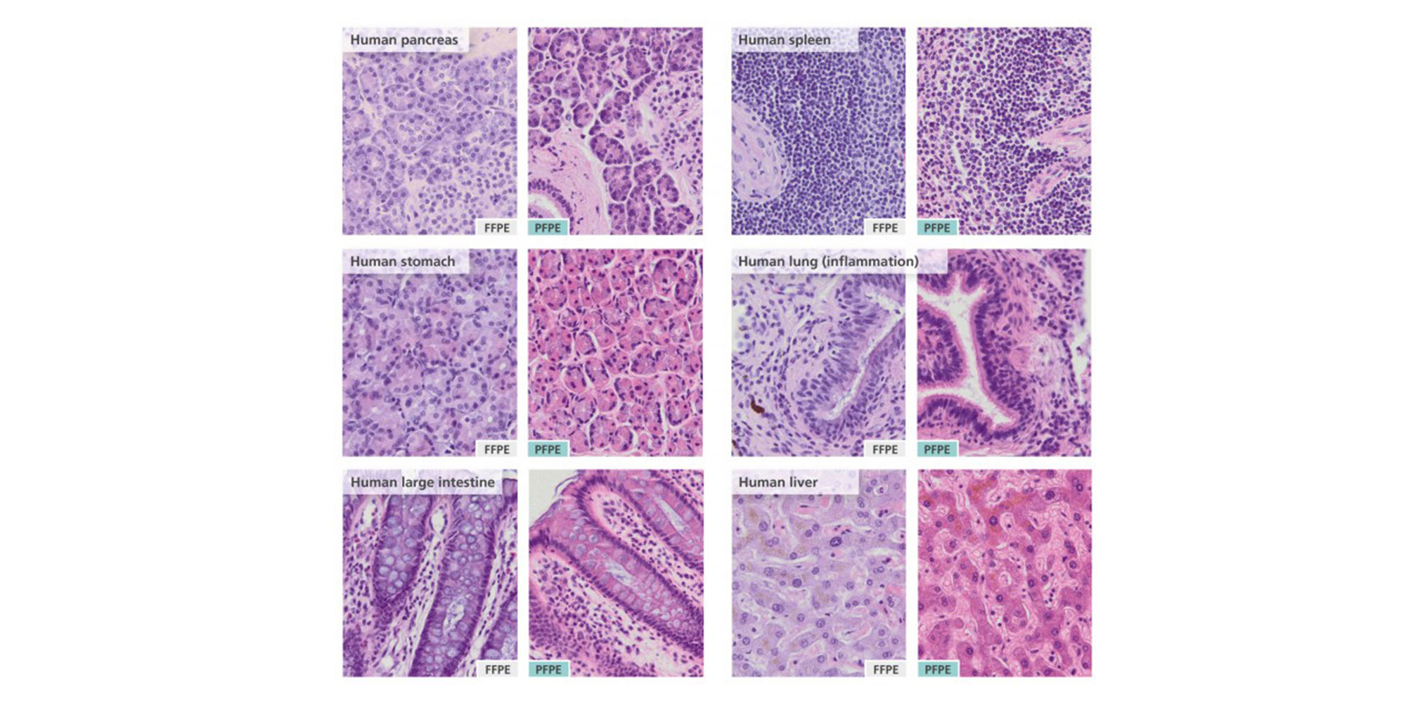
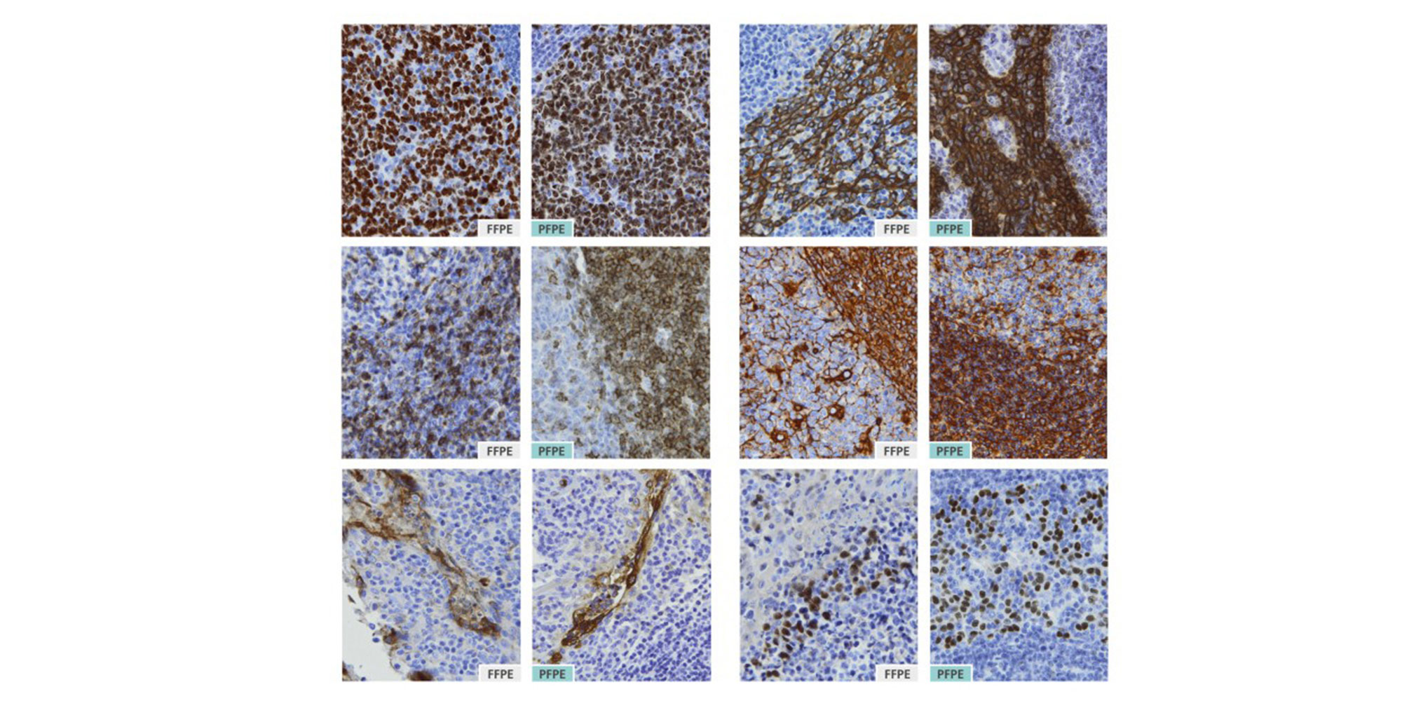
Stabilized samples can be embedded in paraffin for sectioning, and can be stained using standard protocols, such as hematoxylin/eosin (Figure 1) and immunohistochemistry (Figure 2) for tissue sections with high staining intensity and clear morphological features.

Enhanced storage

When tissues are stored in PAXgene Tissue STABILIZER, nucleic acids, proteins and morphology of the tissue sample are stable for up to 7 days at room temperature (15–25°C) or 4 weeks at 2–8°C, depending on the type of tissue. Tissue samples can be stored in the PAXgene Tissue STABILIZER for longer periods at –20°C (–15°C to –30°C) or –80°C (–65°C to –90°C) without negative effects on the morphology of the tissue or the integrity of biomolecules.

Note: Specifications for fixation and storage conditions in PAXgene Tissue FIX and PAXgene Tissue STABILIZER were determined using animal tissues. Storage at 2–8°C for more than 4 weeks must be validated for each tissue type.

Note: For storage at –20°C (–15°C to –30°C) or –80°C (–65°C to –90°C), use cryogenic vials with screw caps filled with PAXgene Tissue STABILIZER. For safety reasons, note that the PAXgene Tissue STABILIZER contains 70% ethanol by volume.

High-quality biomolecule stabilization

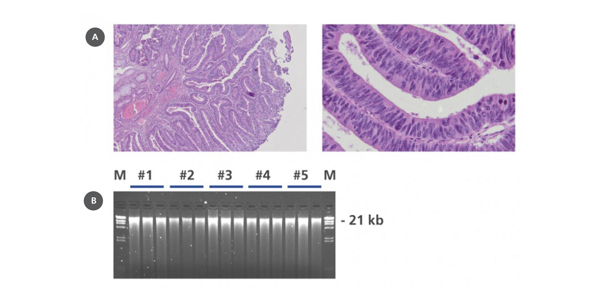
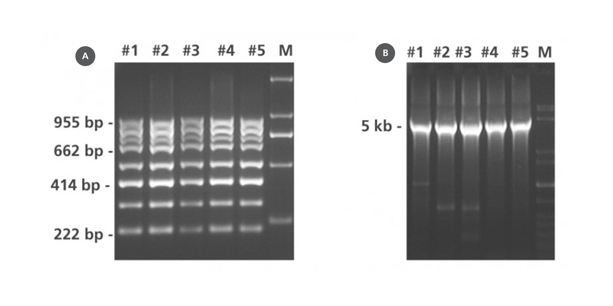
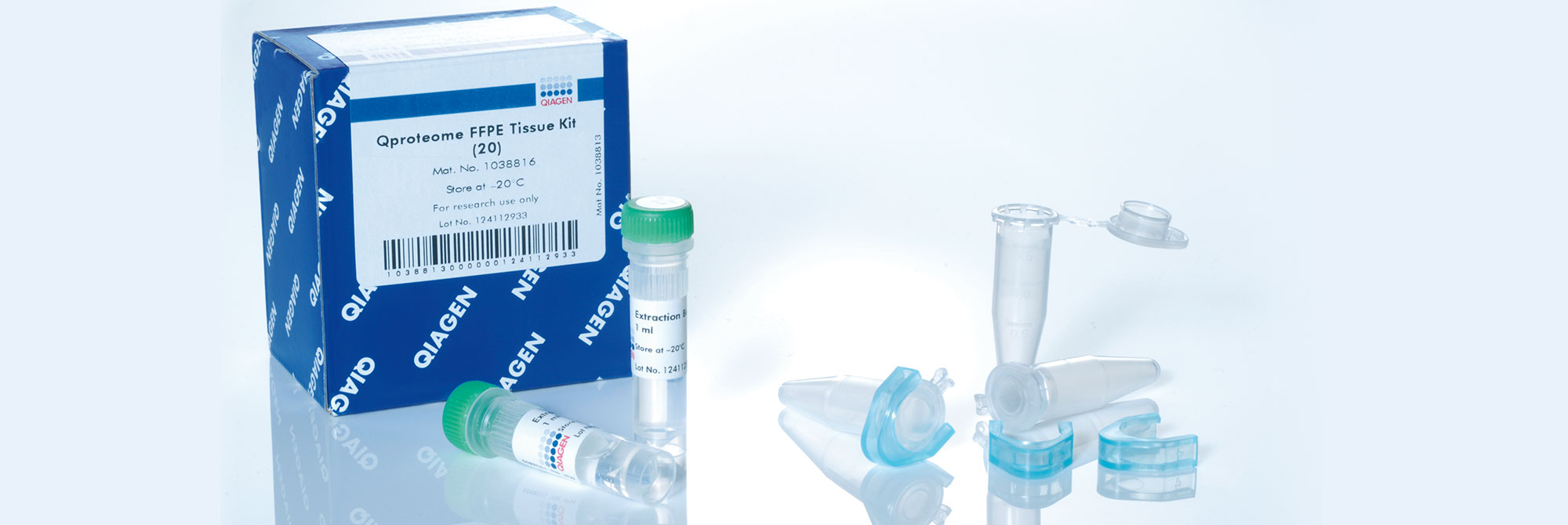
Nucleic acids and proteins of tissues stored in PAXgene Tissue STABILIZER or as PAXgene Tissue-fixed, paraffin-embedded (PFPE) samples are stabilized and can be purified using the corresponding PAXgene Tissue Kit for RNA and miRNA or for DNA. Use the Qproteome FFPE Tissue Kit for purification of proteins. Purified biomolecules are of high quality (Figure 3) and unmodified (Figure 4). The purified biomolecules are highly suited for a range of demanding downstream applications (Figure 5, Figure 6 and Figure 7).

Principle

The methods for tissue fixation currently used in traditional histology are of limited use for molecular analysis. Fixatives that contain formaldehyde crosslink biomolecules and modify nucleic acids and proteins. Such crosslinks lead to nucleic acid degradation during tissue fixation, storage and processing. Since they cannot be removed completely, the resulting chemical modifications can lead to limitations in downstream applications, such as RT-PCR, qPCR and next-generation sequencing. The PAXgene Tissue FIX Container enables formalin-free fixation of tissue specimens. In combination with the PAXgene Tissue STABILIZER Concentrate, which enables formalin-free stabilization, tissue morphology and biomolecule integrity are preserved by avoiding destructive crosslinking and degradation found in formalin-fixed tissues (Figure 9).

Procedure

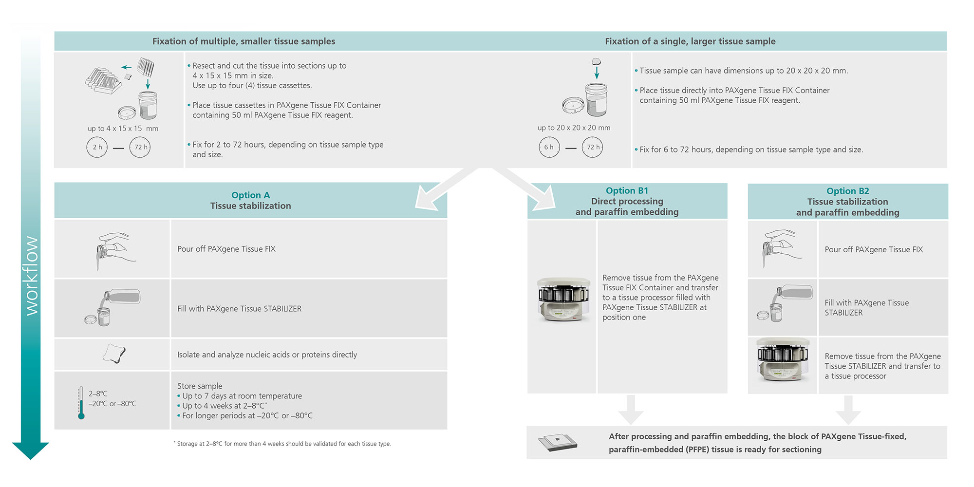
PAXgene Tissue FIX Containers are single-chamber containers prefilled with 50 mL of PAXgene Tissue FIX. PAXgene Tissue FIX Containers can accommodate four standard tissue cassettes (not provided), which can hold tissue samples with a maximum size of 4 x 15 x 15 mm. PAXgene Tissue FIX Containers also offer the possibility for direct fixation of larger tissue samples with a maximum size of 20 x 20 x 20 mm. PAXgene Tissue FIX rapidly penetrates and fixes the tissue. After fixation for 2 to 72 hours, depending on tissue size, PAXgene Tissue FIX is removed and replaced by PAXgene Tissue STABILIZER within the same container. Stabilized samples can be embedded in paraffin for histological studies and nucleic acids and proteins can be isolated from the stabilized samples before or after embedding in paraffin.

Applications

PAXgene Tissue-fixed (PF) and PAXgene Tissue-fixed, paraffin-embedded (PFPE) tissue samples can be used for (see References, Tissue Atlas):

  • Pathological staining, including hematoxylin & eosin (H&E), periodic acid schiff (PAS), resorcin fuchsin, sirius red and Gomori
  • Immunohistochemical staining
  • In situ hybridization

Nucleic acids purified from PF and PFPE tissue samples can be used for demanding downstream applications (see References, Technical Notes), including:

  • RNA and miRNA profiling
  • Long-range and multiplex PCR
  • Next-generation sequencing

Proteins purified from PF and PFPE tissue samples can be used in a range of downstream applications (see References, Technical Notes), including:

  • Western blotting
  • Reverse-phase protein microarrays
  • MALDI imaging mass spectrometry
  • 2-D gel electrophoresis
Human tissue samples were divided into 2 sub-samples. One sub-sample was fixed with PAXgene Tissue FIX and the other half was fixed in neutral-buffered formalin. The fixed tissues were embedded in paraffin, sectioned and stained with hematoxylin and eosin. PFPE: PAXgene Tissue-fixed, paraffin-embedded; FFPE: formalin-fixed, paraffin-embedded.

Figure 1. H&E staining with the PAXgene Tissue System gives results comparable to formalin-fixed tissue.

Human tissue samples were divided into 2 sub-samples. One sub-sample was fixed ...
Human palatine tonsil tissue was fixed in PAXgene Tissue FIX or with neutral-buffered formalin and embedded in paraffin. Primary antibodies to the indicated antigens were linked to a streptavidin-peroxidase conjugate by a biotinylated secondary antibody (LSAB method). Sections were counterstained with hematoxylin. PFPE: PAXgene Tissue-fixed, paraffin-embedded; FFPE: formalin-fixed, paraffin-embedded.

Figure 2. Immunohistochemistry (IHC) staining with the PAXgene Tissue System gives comparable results to formalin-fixed tissue.

Human palatine tonsil tissue was fixed in PAXgene Tissue FIX or with ...
(A) Hematoxylin and eosin (H&E) staining of PAXgene Tissue-fixed, paraffin-embedded (PFPE) human colorectal cancer tissue and (B) DNA on agarose gel electrophoresis using 0.8 % TBE buffered gels with 200 ng genomic DNA isolated in triplicate from 5 cases (#1–5) of human PFPE colorectal cancer. M: markers.

Figure 3. High-quality DNA from PFPE tissue with preserved morphology.

(A) Hematoxylin and eosin (H&E) staining of PAXgene Tissue-fixed, ...
Multiplex and long-range PCR of DNA from PAXgene Tissue-fixed, paraffin-embedded (PFPE) human colorectal cancer tissue (modified according to Viertler et al., (2012) A New technology for stabilization of biomolecules in tissues for combined histological and molecular analyses. J. Mol. Diagn. 14, 458). (A) Multiplex PCR of 8 different genomic DNA fragments ranging from 222 to 955 bp. (B) Long-range PCR of a 5 kb genomic DNA fragment.

Figure 4. DNA without chemical modifications can be used for demanding downstream applications.

Multiplex and long-range PCR of DNA from PAXgene Tissue-fixed, paraffin-embedded ...
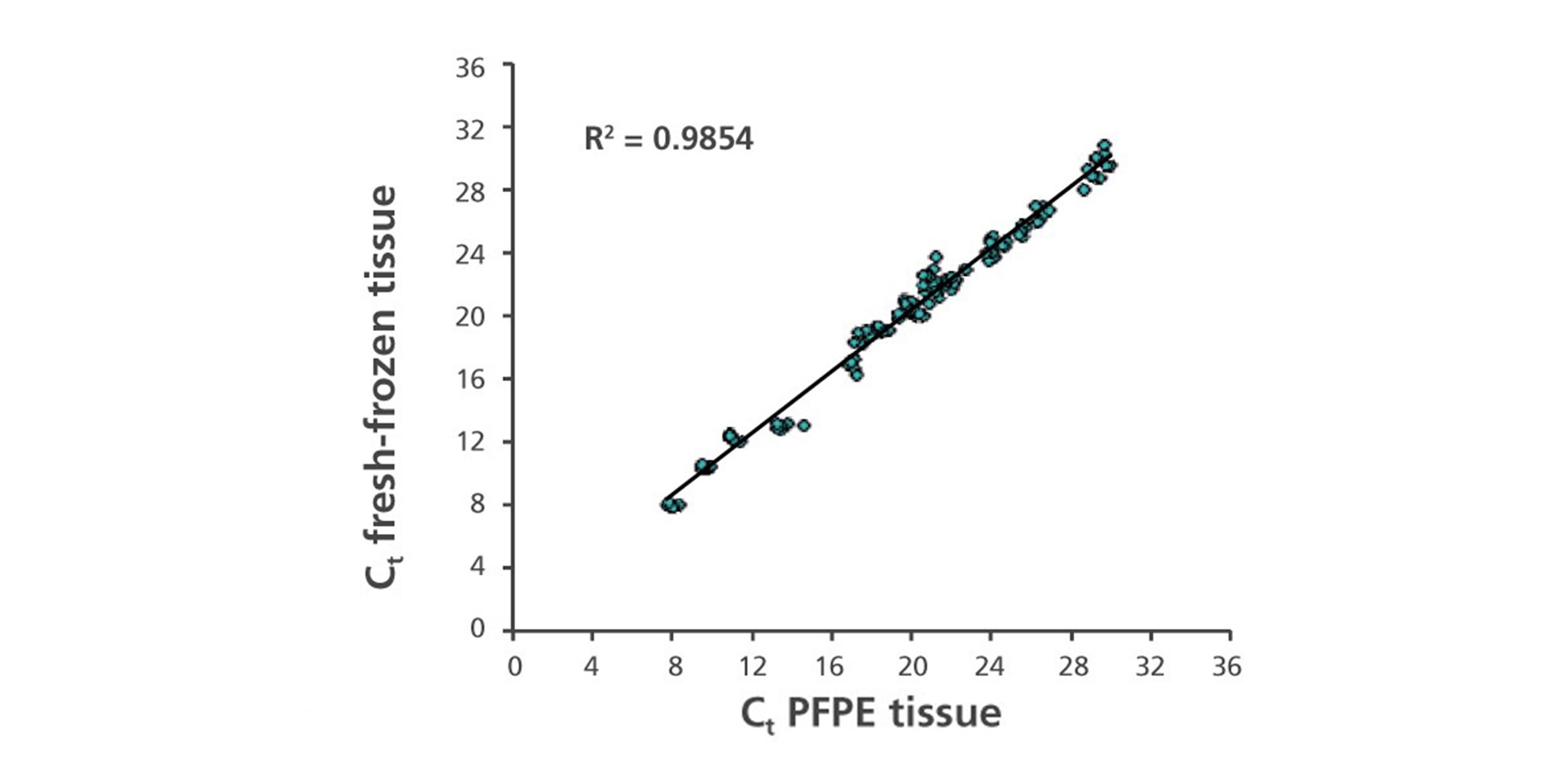
RNA, including miRNA, purified from mirrored human breast cancer tissue fresh frozen in liquid nitrogen using the QIAGEN miRNeasy Kit, or from sections of PAXgene Tissue-fixed, paraffin-embedded (PFPE) tissue using the PAXgene Tissue RNA/miRNA Kit. Shown is a scatterplot of Ct values from different single miRNA-specific RT-qPCR assays using the QIAGEN miScript System: miR9, -10a, -10b, -29a, -103, -125b, -143, -145, -192 and miScript PCR controls RNUA1, RNU5A, RNU6B, SNORD25, SCARNA17, SNORA73A; R2: coefficient of determination.

Figure 5. High concordance of miRNA expression between total RNA isolated from PFPE and fresh-frozen tissue.

RNA, including miRNA, purified from mirrored human breast cancer tissue fresh ...
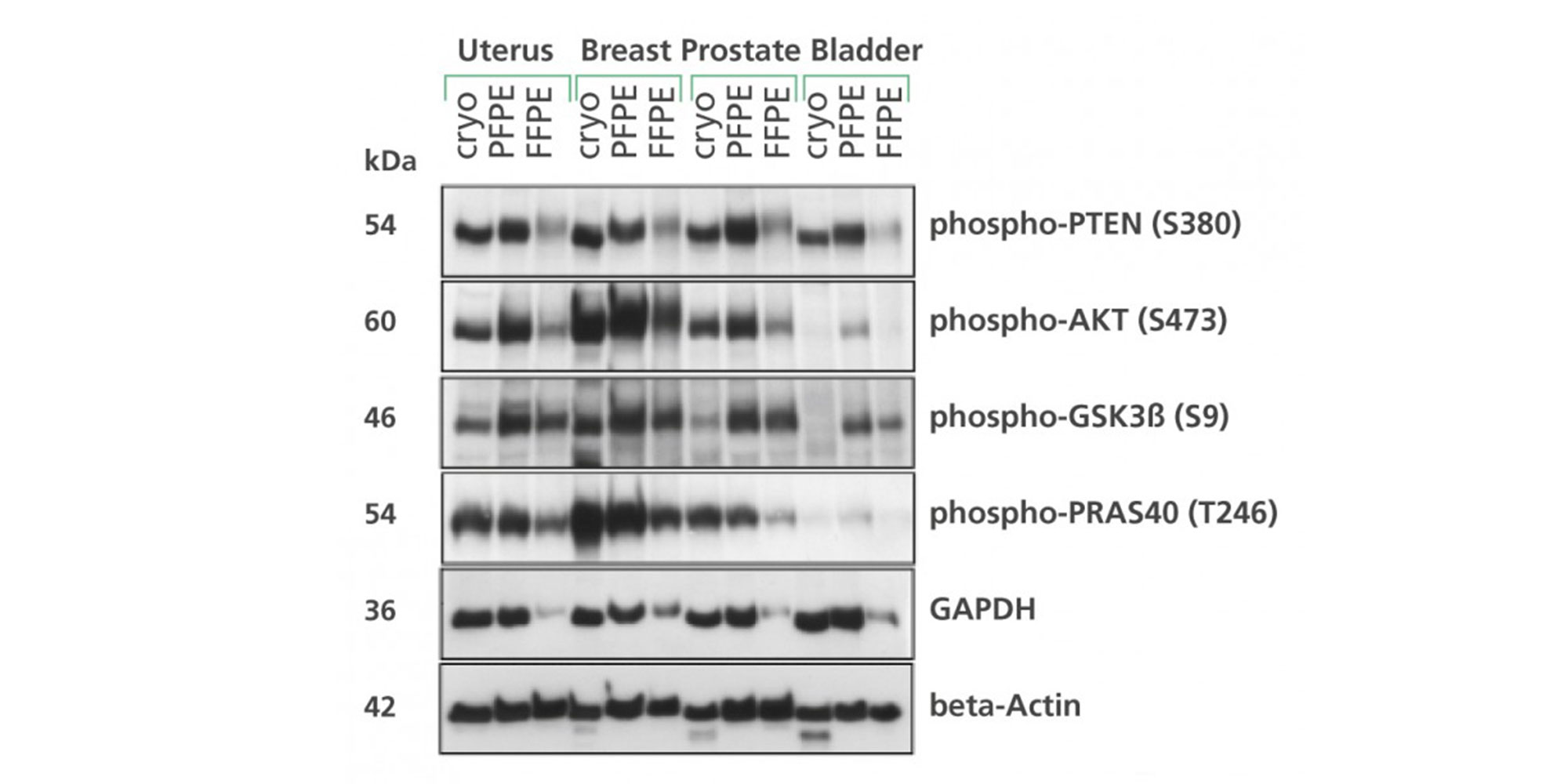
Non-malignant human uterus, breast, prostate and bladder specimens were divided into 3 samples and either flash-frozen in liquid nitrogen (cryo), PAXgene Tissue-fixed, paraffin-embedded (PFPE) or formalin-fixed, paraffin-embedded (FFPE). Proteins from cryo, PFPE, and FFPE tissues were extracted using the extraction buffer EXB Plus (Qproteome FFPE Tissue Kit; described in Ergin et al. 2010, Gündisch et al. 2013 and PAXgene Tissue supplementary protocols). SDS-PAGE and Western blot analysis were performed with 15 μg protein and the indicated antibodies. Data kindly provided by Karl-Friedrich Becker, Technical University of Munich, Germany.

Figure 6. Detection of nondegraded, immunoreactive phosphoproteins from human PFPE tissue.

Non-malignant human uterus, breast, prostate and bladder specimens were divided ...
SYBR Green real-time RT-qPCR was performed with 10 ng RNA from cryopreserved, formalin-fixed, paraffin-embedded (FFPE) or PAXgene Tissue-fixed, paraffin-embedded (PFPE) rat tissue (modified according to Groelz et al. 2013). Depicted are the average delta-Ct values (delta-Ct = Ct[FFPE] – Ct[cryo] or delta-Ct = Ct[PFPE] – Ct[cryo]) from 6 different assays with amplicons ranging from 109 to 465 bp.

Figure 7. RNA purified without chemical modification from PFPE tissue using the PAXgene Tissue RNA/miRNA Kit

SYBR Green real-time RT-qPCR was performed with 10 ng RNA from cryopreserved, ...
Non-malignant human duodenum tissue was divided into 3 samples and either flash-frozen in liquid nitrogen (cryo), or prepared as PFPE or FFPE tissue. Proteins from cryo and PFPE tissue were extracted in 2D buffer (30 mM Tris-HCl pH8.8, 7 M urea, 2 M thiourea, 4 % CHAPS, 75 mM DTT) supplemented with protease inhibitor. Proteins from FFPE tissue were extracted in EXB Plus buffer supplemented with protease inhibitor, precipitated with acetone and resuspended in 2D buffer (as described in Gündisch et al. 2013). Samples (150 μg) were separated by 2-D PAGE. Data kindly provided by Karl-Friedrich Becker, Technical University of Munich, Germany.

Figure 8. Protein extracted from PFPE tissue is suitable for two-dimensional gel electrophoresis.

Non-malignant human duodenum tissue was divided into 3 samples and either ...
Single-chamber container for fixation and stabilization of a single, larger or multiple, smaller tissue samples.

Figure 9. The PAXgene Tissue FIX Container (50 ml) and PAXgene Tissue STABILIZER Workflow.

Single-chamber container for fixation and stabilization of a single, larger or ...
Twelve different PAXgene Tissue-fixed (PF) rat tissue types, including fibrous or fatty, DNA-rich and DNA-poor tissue, processed with the PAXgene Tissue DNA Kit on the QIAcube in one single run. DNA yield per 10 mg tissue.

Figure 10. Yield of high-quality, high molecular weight DNA from 12 different PAXgene Tissue fixed (PF) tissue types, processed on the QIAcube.

Twelve different PAXgene Tissue-fixed (PF) rat tissue types, including fibrous or ...
Twelve different PAXgene Tissue-fixed (PF) rat tissue types, including fibrous or fatty, DNA-rich and DNA-poor tissue, processed with the PAXgene Tissue DNA Kit on the QIAcube in one single run. Ratio of absorbance at 260 and 280 nm.

Figure 11. Absorbance ratio of high-quality, high molecular weight DNA from 12 different PAXgene Tissue fixed (PF) tissue types, processed on the QIAcube.

Twelve different PAXgene Tissue-fixed (PF) rat tissue types, including fibrous or ...
Twelve different PAXgene Tissue-fixed (PF) rat tissue types, including fibrous or fatty, DNA-rich and DNA-poor tissue, processed with the PAXgene Tissue DNA Kit on the QIAcube in one single run. DNA from each tissue type (300 ng) on agarose gel electrophoresis using 1% TBE buffered gels; M: markers.

Figure 12. Agarose gel electrophoresis of high-quality, high molecular weight DNA from 12 different PAXgene Tissue fixed (PF) tissue types, processed on the QIAcube.

Twelve different PAXgene Tissue-fixed (PF) rat tissue types, including fibrous or ...
DNA yield from 3 sections (thickness 10 µm) each of 8 different PAXgene Tissue-fixed, paraffin-embedded (PFPE) rat tissue types processed in triplicate with the PAXgene Tissue DNA Kit on the QIAcube or manually.

Figure 13. Comparison of manual and automated procedure: DNA yield from sections of PAXgene Tissue fixed, paraffin-embedded (PFPE) tissue.

DNA yield from 3 sections (thickness 10 µm) each of 8 different PAXgene ...
Ratio of absorbance at 260 and 280 nm from 3 sections (thickness 10 µm) each of 8 different PAXgene Tissue-fixed, paraffin-embedded (PFPE) rat tissue types processed in triplicate with the PAXgene Tissue DNA Kit on the QIAcube or manually.

Figure 14. Comparison of manual and automated procedure: Absorbance ratio from sections of PAXgene Tissue fixed, paraffin-embedded (PFPE) tissue.

Ratio of absorbance at 260 and 280 nm from 3 sections (thickness 10 µm) each of 8 ...
Agarose gel electrophoresis from 3 sections (thickness 10 µm) each of 8 different PAXgene Tissue-fixed, paraffin-embedded (PFPE) rat tissue types processed in triplicate with the PAXgene Tissue DNA Kit on the QIAcube or manually. DNA from each replicate and tissue type (200 ng) on agarose gel electrophoresis using 1% TBE buffered gels; M: markers.

Figure 15. Comparison of manual and automated procedure: Agarose gel electrophoresis from sections of PAXgene Tissue fixed, paraffin-embedded (PFPE) tissue.

Agarose gel electrophoresis from 3 sections (thickness 10 µm) each of 8 different ...

Resources

(EN) Important Note: PreAnalytiX GmbH street address has changed from “Feldbachstrasse” to “Garstligweg 8”

158.9 KB

Download

Sobin, L. et al. (2024) Histologic and Quality Assessment of Genotype-Tissue Expression (GTEx) Research Samples: A Large Postmortem Tissue Collection. Arch Pathol Lab Med (2024)

Learn more

MP Lemos, RD Astronomo, Y Huang et al. (2024) Enhanced and sustained biodistribution of HIV-1 neutralizing antibody VRC01LS in human genital and rectal mucosa. Nature Communications 2024. Article number: 10332

Learn more

Bravo González-Blas, C. et al. (2024) Single-cell spatial multi-omics and deep learning dissect enhancer-driven gene regulatory networks in liver zonation. Nat Cell Biol 2024

Learn more

Other Products Used in Conjunction


Need Help?

Product Information

For details about our products, reach out 
to our Global Contacts.

Technical Assistance

For troubleshooting or technical inquiries, 
get in touch with Technical Service.

Order Support

For help with placing or tracking an order, 
contact Customer Care.