Select your location:
Image of PAXgene Tissue FIX Container (765312)

PAXgene Tissue FIX Container

For collection, fixation and stabilization of multiple small tissue samples or a single large tissue sample

  • Formalin-free preservation of both morphology and biomolecules
  • Improved molecular results from fixed tissues
  • Tissues can be stored and archived for later processing
  • RNA, miRNA, DNA and/or proteins from one sample

Feature

Specification

Container size

50 ml

Additive

PAXgene Tissue FIX
Note: must be used with PAXgene Tissue STABILIZER Concentrate

Quantity

10 containers

Specimen size

  • 4 tissue cassettes: up to 4 x 15 x 15 mm or
  • 1 tissue sample up to 20 x 20 x 20 mm

Fixation time in PAXgene Tissue FIX Container

2–72 h, depending on tissue sample type and size

Length of stabilization in PAXgene Tissue STABILIZER Concentrate

  • up to 7 days at room temperature (15–25°C)
  • up to 4 weeks at 2–8°C*
  • long-term at –20°C or –80°C**

Archiving options for PFPE***

  • short-term, refrigerated at 5°C (2–8°C)
  • ideally, frozen at –20°C (–15°C to –30°C)**

*   Storage at 2–8°C for more than 4 weeks
    must be validated for each tissue type

**  Long-term storage studies are ongoing
*** PAXgene Tissue-fixed, paraffin-embedded


Intended Use

The PAXgene Tissue FIX Container is intended for the collection, fixation and stabilization of tissue specimens. The container is intended to be used as part of the PAXgene Tissue System and must be used in conjuction with the PAXgene Tissue STABILIZER Concentrate (see Procedure below). The PAXgene Tissue System additionally includes the PAXgene Tissue RNA/miRNA Kit for the isolation of total RNA, including miRNA, and PAXgene Tissue DNA Kit for DNA. Supplementary protocols are available for other applications, including purification of proteins.

For research use only. Not for use in diagnostic procedures. No claim or representation is intended to provide information for the diagnosis, prevention, or treatment of a disease. The performance characteristics of this product have not been fully established.

Order information

Product

Catalog No.

Price

PAXgene TissueFIX Cont. (50 ml) Kit

For fixation and stabilization of tissue specimen: 10 prefilled Reagent Containers containing 50 ml of PAXgene Tissue FIX to be used in conjunction with PAXgene Tissue STABILIZER Reagent.

765312
225 USD
Order now
Order now

Details

Performance

PAXgene Tissue FIX Containers are 50 ml containers prefilled with PAXgene Tissue FIX, which rapidly penetrates and fixes tissues, preserving tissue morphology and biomolecules. The container accommodates up to 4 standard tissue cassettes or a single, large tissue specimen. Fixation with PAXgene Tissue FIX is comparable to formalin fixation, but avoids destructive nucleic acid and protein crosslinking and degradation. After fixation, PAXgene Tissue FIX is replaced with 50 ml PAXgene Tissue STABILIZER Concentrate in the same container.

Note: Fixation rate depends on tissue type and size.

Preserved morphology

Stabilized samples can be embedded in paraffin for sectioning, and can be stained using standard protocols, such as hematoxylin/eosin (Figure 1) and immunohistochemistry (Figure 2) for tissue sections with high staining intensity and clear morphological features.

Enhanced storage

When tissues are stored in PAXgene Tissue STABILIZER, nucleic acids, proteins and morphology of the tissue sample are stable for up to 7 days at room temperature (15–25°C) or 4 weeks at 2–8°C, depending on the type of tissue. Tissue samples can be stored in the PAXgene Tissue STABILIZER for longer periods at –20°C (–15°C to –30°C) or –80°C (–65°C to –90°C) without negative effects on the morphology of the tissue or the integrity of biomolecules.

Note: Specifications for fixation and storage conditions in PAXgene Tissue FIX and PAXgene Tissue STABILIZER were determined using animal tissues. Storage at 2–8°C for more than 4 weeks must be validated for each tissue type.

Note: For storage at –20°C (–15°C to –30°C) or –80°C (–65°C to –90°C), use cryogenic vials with screw caps filled with PAXgene Tissue STABILIZER. For safety reasons, note that the PAXgene Tissue STABILIZER contains 70% ethanol by volume.

High-quality biomolecule stabilization

Nucleic acids and proteins of tissues stored in PAXgene Tissue STABILIZER or as PAXgene Tissue-fixed, paraffin-embedded (PFPE) samples are stabilized and can be purified using the corresponding PAXgene Tissue Kit for RNA and miRNA or for DNA. Use the Qproteome FFPE Tissue Kit for purification of proteins. Purified biomolecules are of high quality (Figure 3) and unmodified (Figure 4). The purified biomolecules are highly suited for a range of demanding downstream applications (Figure 5, Figure 6 and Figure 7).

Principle

The methods for tissue fixation currently used in traditional histology are of limited use for molecular analysis. Fixatives that contain formaldehyde crosslink biomolecules and modify nucleic acids and proteins. Such crosslinks lead to nucleic acid degradation during tissue fixation, storage and processing. Since they cannot be removed completely, the resulting chemical modifications can lead to limitations in downstream applications, such as RT-PCR, qPCR and next-generation sequencing. The PAXgene Tissue FIX Container enables formalin-free fixation of tissue specimens. In combination with the PAXgene Tissue STABILIZER Concentrate, which enables formalin-free stabilization, tissue morphology and biomolecule integrity are preserved by avoiding destructive crosslinking and degradation found in formalin-fixed tissues (Figure 9).

Procedure

PAXgene Tissue FIX Containers are single-chamber containers prefilled with 50 ml of PAXgene Tissue FIX. PAXgene Tissue FIX Containers can accommodate four standard tissue cassettes (not provided), which can hold tissue samples with a maximum size of 4 x 15 x 15 mm. PAXgene Tissue FIX Containers also offer the possibility for direct fixation of larger tissue samples with a maximum size of 20 x 20 x 20 mm. PAXgene Tissue FIX rapidly penetrates and fixes the tissue. After fixation for 2 to 72 hours, depending on tissue size, PAXgene Tissue FIX is removed and replaced by PAXgene Tissue STABILIZER within the same container. Stabilized samples can be embedded in paraffin for histological studies and nucleic acids and proteins can be isolated from the stabilized samples before or after embedding in paraffin.

Applications

PAXgene Tissue-fixed (PF) and PAXgene Tissue-fixed, paraffin-embedded (PFPE) tissue samples can be used for (see References, Tissue Atlas):

  • Pathological staining, including hematoxylin & eosin (H&E), periodic acid schiff (PAS), resorcin fuchsin, sirius red and Gomori
  • Immunohistochemical staining
  • In situ hybridization

Nucleic acids purified from PF and PFPE tissue samples can be used for demanding downstream applications (see References, Technical Notes), including:

  • RNA and miRNA profiling
  • Long-range and multiplex PCR
  • Next-generation sequencing

Proteins purified from PF and PFPE tissue samples can be used in a range of downstream applications (see References, Technical Notes), including:

  • Western blotting
  • Reverse-phase protein microarrays
  • MALDI imaging mass spectrometry
  • 2-D gel electrophoresis

Resources

PAXgene TIssue FIX Container Handbook

638.4 KB

Download

PAXgene Tissue STABILIZER Concentrate (150 ml) Handbook

181.3 KB

Download

Deparaffinization of PAXgene Tissue-fixed, Paraffin-embedded Tissue (PFPE) Sections with Deparaffinization Solution

614.1 KB

Download

Purification of Genomic DNA from Microdissected PAXgene Tissue-fixed, Paraffin-embedded (PFPE) and PAXgene Tissue-fixed, Cryo-embedded (PFCE) Tissues

699.3 KB

Download

Purification of Genomic DNA from Sections of PAXgene Tissue-fixed, Cryo-embedded (PFCE) Tissue Placed Directly Into a Microcentrifuge Tube

589.0 KB

Download

Purification of Total RNA, including miRNA, from Sections of PAXgene Tissue-fixed, Cryo-embedded (PFCE) Tissue Placed Directly into a Microcentrifuge Tube

606.8 KB

Download

Purification of Total RNA, including miRNA, from Microdissected PAXgene Tissue-fixed, Paraffin-embedded (PFPE) and PAXgene Tissue-fixed, Cryo-embedded (PFCE) Tissues

662.9 KB

Download

Preparation of Sections from PAXgene Tissue-fixed, Paraffin-embedded (PFPE) and PAXgene Tissue-fixed, Cryo-embedded (PFCE) Tissues for Manual or Laser Microdissection (LMD)

608.8 KB

Download

Preparation of PFPE Tissue Sections for Use with In Situ Hybridization (ISH) Staining Assays

591.7 KB

Download

Purification of Full-length Proteins from PAXgene Tissue-fixed and Stabilized (PF) Tissue Samples

614.2 KB

Download

Purification of Full-length Proteins from Sections of PAXgene Tissue-fixed, Cryo-embedded (PFCE) Tissue

635.8 KB

Download

Purification of Full-length Proteins from Sections of PAXgene Tissue-fixe, Paraffin-embedded (PFPE) Tissue

636.4 KB

Download

Simultaneous Purification of Genomic DNA and Total RNA, including miRNA, from Sections of PAXgene Tissue-fixed, Paraffin-embedded (PFPE) Tissue

82.6 KB

Download

Manual Processing of Tissue Specimens Treated with the PAXgene Tissue System

334.1 KB

Download

Cryo-embedding Tissue Specimens Fixed and Stabilized with the PAXgene Tissue System

1.1 MB

Download

PAXgene Tissue System Brochure

1.9 MB

Download

DNA Isolation with the PAXgene Tissue DNA Kit

1.9 MB

Download

Protein extraction from PFPE

573.4 KB

Download

Hematoxylin and Eosin (H&E) Staining with the PAXgene Tissue System

6.5 MB

Download

(EN) Important Note: PreAnalytiX GmbH street address has changed from “Feldbachstrasse” to “Garstligweg 8”

158.9 KB

Download

Morphology and RNA preservation in PAXgene Tissue-fixed, paraffin-embedded tissue (PFPE) stored for 18 months at different temperatures

955.8 KB

Download

RNA stability in tissue samples, fixed and stabilized with the PAXgene Tissue System, for up to 7 days at 22°C or 2 months at 4°C

329.8 KB

Download

Effect of epitope retrieval conditions on immunohistochemical staining of PFPE tonsil tissue with anti-human Ki-67 antigen (clone MIB-1)

2.4 MB

Download

Detection of PI3K mutational status in DNA from human breast cancer PFPE tissue using the PI3K Mutation Test Kit (QIAGEN®)

1.2 MB

Download

Effect of Epitope Retrieval Conditions on Immunohistochemical Staining of PFPE Tonsil Tissue with Anti-human Ki-67 Antigen (Clone MIB-1)

724.5 KB

Download

Vacuum Sealing of Fixed and Stabilized Tissue with a FoodSaver Vaccum Sealer for Dry and Safe Transport

864.9 KB

Download

Influence of Formalin Contamination During Processing of PAXgene Tissue-fixed, Paraffin-embedded Tissue (PFPE) on RNA Yield, Integrity and Performance in Quantitative RT-PCR

661.1 KB

Download

Influence on RNA Yield and Integrity of Modifications to the Processing Protocol for PAXgene Tissue-fixed, Paraffin-embedded (PFPE) Rat Tissue

633.0 KB

Download

Yield, Purity and Integrity of RNA Purified from PAXgene Tissue-fixed, Paraffin-embedded (PFPE) Rat Tissue

603.8 KB

Download

Quantitative Analysis of KRAS and BRAF Mutational Status in DNA from PAXgene Tissue-fixed, Paraffin-embedded (PFPE) Tissue Using Pyrosequencing Technology

1.3 MB

Download

Simultaneous Preservation of RNA and Morphology in Tissue Samples Fixed with PAXgene Tissue Fix and Stored in PAXgene Tissue STABILIZER at –20°C or –80°C

1.3 MB

Download

RNA Stability in TIssue Samples, Fixed and Stabilized with the PAXgene Tissue System, for up to 7 Days at 22°C or 2 Months at 4°C

524.9 KB

Download

Detection of PI3K Mutational Status in DNA from Human Breast Cancer PFPE Tissue Using the PI3K Mutation Test Kit (QIAGEN)

1,004.0 KB

Download

Morphology and RNA Preservation in PAXgene Tissue-fixed, Paraffin-embedded Tissue (PFPE) Stored at 18 Months at Different Temperatures

483.8 KB

Download

MSDS PAXgene Tissue FIX Container (10 x 50 ml)

Learn more

Storage of Tissue in PAXgene Tissue STABILIZER: RNA and Morphology Preservation after 5 Years at –20 and 3 Years at –80°C (Groelz 2014)

887.7 KB

Download

PAXgene; a Beneficial Formalin Alternative to Study Lung Cancer (Southwood 2018)

2.1 MB

Download

Implementation of Formalin-free PAXgene Tissue Fixative into Routine Use: Evaluation of H&E Morphology, IHC and FISH (Meecham 2018)

1.7 MB

Download

RT-PCR Performance of RNA Obtained From Archived Formalin or PAXgene Tissue Fixed, Paraffin-embedded (FFPE and PFPE) Blocks of Tissue (Groelz 2014)

7.2 MB

Download

Stability of Nucleic Acids in Archived Formalin and PAXgene Tissue Fixed, Paraffin-embedded (FFPE and PFPE) Blocks of Tissue (Groelz 2014)

2.5 MB

Download

Preservation of Morphology and Biomolecules Within Tissue Stored for Three Years at –80°C in PAXgene Tissue Stabilizer Reagent (Groelz 2012)

890.5 KB

Download

PAXgene vs. Formalin Fixed Tissue: A Comparison of Tissue Morphology and RNA Quality (Groelz 2012)

5.0 MB

Download

PAXgene Tissue Fixation Technology for Simultaneous Preservation of Morphology and Biomolecules (Groelz 2012)

1.2 MB

Download

DNA Quality Measurement and Somatic Mutation Profiling in PAXgene Tissue Samples with qBiomarker Somatic Mutation PCR Arrays (Long 2012)

1.2 MB

Download

Evaluation of the PAXgene Tissue System: Preservation of Morphology and Gene Expression in Human Melanoma (Hesse 2011)

973.5 KB

Download

PAXgene Tissue: A New TIssue Fixation Technology for Simultaneous Preservation of Morphology and Nucleic Acids (Groelz 2011)

542.1 KB

Download

A New Tissue Fixative for Biomarker Discovery: Gene Expression and miRNA in PAXgene Tissue-fixed, Paraffin-embedded (PFPE) Colorectal Cancer (CRC) and Breast Cancer Tissue vs. Formalin-fixed, Paraffin-embedded (FFPE) Tissue (Groelz 2011)

413.4 KB

Download

Preservation of Gene Expression Profile and Histomorphology in Human Breast Tumor Tissue with the New PAXgene Tissue System (Groelz 2010)

750.8 KB

Download

Morphological, Epigenomic and Mutational Analyses of PAXgene Tissue-fixed, Paraffin-embedded (PFPE) Colorectal Cancer (CRC) Specimens — Comparison to Formalin-fixed, Paraffin-embedded (FFPE) and Snap-frozen Samples (Groelz 2010)

1.1 MB

Download

Can we preserve morphology and nucleic acid integrity for molecular pathology? (Mathieson 2010)

728.1 KB

Download

Long-term Storage of TIssue Specimens at –20°C to –80°C with Preservation of Morphology and Nucleic Acids Within Frozen Tissue (Groelz 2009)

676.9 KB

Download

Preservation of Histomorphology and Nucleic Acids in Human Breast Tumor TIssue with the New PAXgene Tissue System — A Study with Comparison to Formalin Fixation (Groelz 2009)

934.1 KB

Download

New Fixation Technology for Simultaneous Preservation of Mophology and Nucleic Acids in Tissue (Groelz 2008)

516.6 KB

Download

Bravo González-Blas, C. et al. (2024) Single-cell spatial multi-omics and deep learning dissect enhancer-driven gene regulatory networks in liver zonation. Nat Cell Biol 2024

Learn more

Nurminen et al.(2023) Cancer origin tracing and timing in two high-risk prostate cancers using multisample whole genome analysis: prospects for personalized medicine. Genome Medicine volume 15, Article number: 82

Learn more

Takamatsu, R. et al. (2023) Clinical predominance of whole‐exome sequencing to evaluate microsatellite instability status. Cancer Science. 2023;00:1–12

Learn more

R, Li. et al. (2022) Mapping single-cell transcriptomes in the intra-tumoral and associated territories of kidney cancer. Cancer Cell. 2022-Elsevier

Learn more

Karras, P. et al. (2022) A cellular hierarchy in melanoma uncouples growth and metastasis, Nature 2022

Learn more

Radani, N. et al. (2022) Analysis of Fecal, Salivary, and Tissue Microbiome in Barrett's Esophagus, Dysplasia, and Esophageal Adenocarcinoma. Gastro Hep Advances 2022;1:755–766

Learn more

Marsden, T. et al. (2022) The ReIMAGINE prostate cancer risk study protocol: A prospective cohort study in men with a suspicion of prostate cancer who are referred onto an MRI-based diagnostic pathway with donation of tissue, blood and urine for biomarker analyses. PLoS One. 2022; 17(2): e0259672.

Learn more

Hingorani, D. et al. (2022) Monomethyl auristatin antibody and peptide drug conjugates for trimodal cancer chemo-radio-immunotherapy. Nat Commun. 2022; 13: 3869.

Learn more

Meecham, A. et al. (2021) Alternative tissue fixation for combined histopathological and molecular analysis in a clinically representative setting. Histochem Cell Biol.

Learn more

Robinson, P. S. et al. (2021) Increased somatic mutation burdens in normal human cells due to defective DNA polymerases. Nat Genet.

Learn more

Lahiri, P. et al. (2021) Comprehensive Evaluation of PAXgene Fixation on Oral Cancer Tissues Using Routine Histology, Immunohistochemistry, and FTIR Microspectroscopy. Biomolecules 2021, 11, 889.

Learn more

Garcia Frasquilho, S. et al. (2021) Do Tissues Fixed in a Non-cross-linking Fixative Require a Dedicated Formalin-free Processor? Journal of Histochemistry & Cytochemistry 1–17.

Learn more

Spencer Chapman, M. et al. (2021) Lineage tracing of human development through somatic mutations. Nature.com.

Learn more

Grossmann, S. et al (2021) Development, maturation, and maintenance of human prostate inferred from somatic mutations. Cell Stem Cell, Volume 28, Issue 7, Pages 1262-1274.e5.

Learn more

Ellis, P. et al. (2021) Reliable detection of somatic mutations in solid tissues by laser-capture microdissection and low-input DNA sequencing. PubMed.gov.

Learn more

Wiethaler, M. et al. (2019) BarrettNET – a prospective registry for risk estimation of patients with Barrett's esophagus to progress to adenocarcinoma. Diseases of the Esophagus, Volume 32, Issue 8.

Learn more

Stumptner, C., Pabst, D.,Loibner, M., Viertler, C., Zatloukal, K. (2019) The impact of crosslinking and non-crosslinking fixatives on antigen retrieval and immunohistochemistry. N. Biotechnol. 52: 69.

Learn more

Högnäs, G. et al. (2018) Feasibility of Prostate PAXgene Fixation for Molecular Research and Diagnostic Surgical Pathology: Comparison of Matched Fresh Frozen, FFPE, and PFPE Tissues. Am. J. Surg

Learn more

Urban, C. et al. (2018) PAXgene fixation enables comprehensive metabolomic and proteomic analyses of tissue specimens by MALDI MSI. Biochim. Biophys. Acta Gen. Subj. 1862, 51. E-published in 2017

Learn more

Yamazaki, M. et al. (2018) PAXgene-fixed paraffin-embedded sample is applicable to laser capture microdissection with well-balanced RNA quality and tissue morphology. J. Toxicol. Pathol. 31: 213.

Learn more

Liu, Y. and Edward, D.P. (2017) Assessment of PAXgene Fixation on Preservation of Morphology and Nucleic Acids in Microdissected Retina Tissue. Curr. Eye Res. 42, 104. E-published in 2016. doi: 0.

Learn more

GTEx Consortium et al. (2017) Genetic effects on gene expression across human tissues. Nature. 550, 204. doi: 10.1038/nature24277

Learn more

Ruusuvuori, P. et al. (2016) Feature-based analysis of mouse prostatic intraepithelial neoplasia in histological tissue sections. J. Pathol. Inform. 7, 5. doi: 10.4103/2153-3539.175378

Learn more

Loibner, M. et al. (2016) Pathogen Inactivating Properties and Increased Sensitivity in Molecular Diagnostics by PAXgene, a Novel Non-Crosslinking Tissue Fixative. PLoS One 11, e0151383. doi: 10.

Learn more

Mathieson, W. et al. (2016) A Critical Evaluation of the PAXgene Tissue Fixation System:  Morphology, Immunohistochemistry, Molecular Biology, and Proteomics. Am. J. Clin. Pathol. 146, 25. doi: 0

Learn more

Oberauner-Wappis, L. et al. (2016) Protocol for HER2 FISH determination on PAXgene-fixed and paraffin-embedded tissue in breast cancer. Int. J. Exp. Pathol. 97, 202. doi: 10.1111/iep.12185

Learn more

Korenkova, V. et al. (2016) The focus on sample quality: Influence of colon tissue collection on reliability of qPCR data. Sci. Rep. 6, 29023. doi: 10.1038/srep29023

Learn more

Yamaguchi, T. et al. (2015) Comprehensive DNA Methylation and Extensive Mutation Analyses of HER2-Positive Breast Cancer. Oncology 88, 377. doi: 0.1159/000369904

Learn more

Carithers, L.J. et al. (2015) A Novel Approach to High-Quality Postmortem Tissue Procurement: The GTEx Project. Biopreserv. Biobank. 13, 311. doi: 10.1089/bio.2015.0032

Learn more

Taavela, J. et al. (2019) Histological, immunohistochemical and mRNA gene expression responses in coeliac disease patients challenged with gluten using PAXgene fixed paraffin-embedded duodenal biopsies. BMC Gastroenterol 19, 189 (2019).

Learn more

Gillard, M. et al. (2015) Next-gen tissue: preservation of molecular and morphological fidelity in prostate tissue. Am. J. Transl. Res. 7, 1227.

Learn more

Hara, K. et al. (2015) Surgical Specimens of Colorectal Cancer Fixed with PAXgene Tissue System Preserve High-Quality RNA. Biopreserv. Biobank. 13, 325. doi: 10.1089/bio.2014.0101

Learn more

Gündisch, S. et al. (2014) Evaluation of colon cancer histomorphology: a comparison between formalin and PAXgene tissue fixation by an international ring trial. Virchows Arch. 465, 509. doi: 10.1

Learn more

Groelz, D. et al. (2013) Non-Formalin Fixative versus Formalin-fixed Tissue: A Comparison of Histology and RNA Quality. Exp. Mol. Pathol. 94, 188. doi: 10.1016/j.yexmp.2012.07.002

Learn more

Gündisch, S. et al. (2013) The PAXgene Tissue System Preserves Phosphoproteins in Human Tissue Specimens and Enables Comprehensive Protein Biomarker Research. PLoS One 8, e60638. doi: 10.1371/jou

Learn more

Lonsdale, J. et al. (2013) The Genotype-Tissue Expression (GTEx) project. Nature Genetics 45, 580. doi: 0.1038/ng.2653

Learn more

Kap, M. et al. (2013) Inactivation of Influenza A virus, Adenovirus, and Cytomegalovirus with PAXgene Tissue Fixative and Formalin. Biopreserv. Biobank. 11, 229. doi: 10.1089/bio.2013.0010

Learn more

Staff, S. et al. (2013) Preservation of nucleic acids and tissue morphology in paraffin-embedded clinical samples: comparison of five molecular fixatives. J. Clin. Pathol. 66, 807. doi: 10.1136/jc

Learn more

Baker, M. (2012) Biorepositories: Building better biobanks. Nature 486, 141. doi: 10.1038/486141a

Learn more

Viertler, C. et al. (2012) A New Technology for Stabilization of Biomolecules in Tissues for Combined Histological and Molecular Analyses. J. Mol. Diagn. 14, 458. doi: 0.1016/j.jmoldx.2012.05.002

Learn more

Kap, M. et al. (2011) Histological Assessment of PAXgene Tissue Fixation and Stabilization Reagents. PLoS ONE 6, e27704. doi: 10.1371/journal.pone.0027704

Learn more

Ergin, B. et al. (2010) Proteomic analysis of PAXgene-fixed tissues. J. Proteome Res. 9, 5188. doi: 10.1021/pr100664e

Learn more

Groelz, D. et al. (2018) Impact of storage conditions on the quality of nucleic acids in paraffin embedded tissues. PLoS ONE 13(9): e0203608.

Learn more

PAXgene Tissue FIX Container

Tissue fixation and stabilization chemistry


1. Which fixation method is used in the PAXgene Tissue System?


The PAXgene Tissue System uses an acidic and alcoholic fixation without formalin that does not result in crosslinking of biomolecules.

2. What is the composition of PAXgene Tissue FIX?
The PAXgene Tissue FIX fixation reagent contains alcohols and an acid, in addition to other stabilization agents.

3. What is the composition of PAXgene Tissue STABILIZER?
The PAXgene Tissue STABILIZER stabilization reagent contains alcohol and other stabilization agents. It is available in bulk as a concentrate.

4. Are the two reagents used in the PAXgene Tissue System obligatory?
The PAXgene Tissue System involves two processes: fixation and stabilization. PAXgene Tissue FIX provides rapid penetration and fixation that effectively stops all enzymatic activity throughout the tissue. The tissue can remain in the fixative for maximum 72 h. For long-term transportation and storage, PAXgene Tissue STABILIZER stops fixation and stabilizes the specimen.

Tissue fixation and stabilization

1. What is the maximum tissue size that can be fixed in a PAXgene Tissue FIX Container (50 ml)?
Up to 4 standard tissue cassettes, each containing tissue samples with a maximum size of 4 x 15 x15 mm, or alternatively, a single tissue sample with a maximum size of 20 x 20 x 20 mm can be placed into a PAXgene Tissue FIX Container. If using a larger tissue sample surrounded by fat (e.g., from a lymph node) or a capsule (e.g., from kidney, liver or spleen), partially cut into the tissue every 5 mm (lamination) to enhance permeation of the fixation reagent. If samples are larger than recommended, the fast and even penetration of fixation reagent is compromised. This may result in quality reduction of tissue morphology and integrity of nucleic acids.

2. How long is the fixation time?
Depending on tissue size specimen(s) must be fixed at room temperature (15–25°C) for a minimum of 2 h (for samples up to 4 x 15 x 15 mm), or for a minimum of 6 h (for samples up to 20 x 20 x 20 mm). Fixation should be stopped by transfer into PAXgene Tissue STABILIZER after a maximum of 72 hours fixation.

For biopsies with a thickness of 1 mm or less, fixation time can be reduced to 30–60 min.

3. Which conditions are recommended for the storage of tissues in PAXgene Tissue STABILIZER?
Depending on tissue type, standard storage conditions in PAXgene Tissue STABILIZER are up to 7 days at room temperature (15–25°C) or up to 4 weeks at 2–8°C. Storage at 2–8°C for more than 4 weeks must be validated for each tissue type. For longer storage, samples can be kept at–15°C to –30°C or –65°C to –90°C. Long-term storage studies are ongoing. For the latest results, see the poster "RNA and Morphology Preservation after 5 years at –20°C and 3 years at –80°C" under Resources.

4. Are PAXgene Tissue FIX Containers suitable for long-term storage at freezing temperature?
No. For storage at –15°C to –30°C or –65°C to –90°C use cryogenic vials with screw caps filled with PAXgene Tissue STABILIZER. For safety reasons, note that the PAXgene Tissue STABILIZER contains 70% ethanol.

Processing

1. Is it possible to use a standard processor – the kind used routinely for formalin-fixed samples – for dehydration and paraffin infiltration of PAXgene Tissue-treated samples?
Yes. All processors commonly used for formalin-fixed samples can be used to produce PAXgene Tissue-fixed, paraffin-embedded (PFPE) blocks of tissue.

We recommend keeping alcohol for processing PAXgene Tissue-treated samples separate from alcohol used for processing formalin-fixed samples for at least the first five positions/steps in the processing. With this precaution, it is possible to process PAXgene Tissue-fixed and formalin-fixed samples on the same instrument.

2. Is it possible to process formalin-fixed and PAXgene Tissue-fixed samples together in one run?
Parallel processing of formalin-fixed and PAXgene Tissue-fixed samples can lead to reduction of nucleic acid yield and integrity from PFPE samples through formalin contamination of reagents.

3. Is it necessary to clean a processor normally used for formalin-fixed tissue before using it with PAXgene Tissue-fixed tissue?
No. Special cleaning is not required. However, when processing PAXgene Tissue-treated specimens, do not use reagents contaminated with formalin. Residual formalin can lead to significant reduction of nucleic acid yield and integrity from PFPE tissue samples (see Technical Note "Influence of formalin contamination during processing of PAXgene Tissue fixed, paraffin-embedded tissue (PFPE) on RNA yield, integrity, and performance in quantitiative RT-PCR"). We recommend keeping alcohol for processing PAXgene Tissue-treated samples separate from alcohol used for processing formalin-fixed samples for at least the first five positions/steps in the processing. With this precaution, it is possible to process PAXgene Tissue-fixed and formalin-fixed samples on the same instrument.

4. Is a special processing protocol needed for the PAXgene Tissue System?
To prevent biomolecule degradation during processing, dehydration must start with at least 70–100% ethanol. We recommed using low-melting paraffin (melting point ≤56°C) and incubation in liquid paraffin for no longer than 3 hours.

Processing protocols for the PAXgene Tissue System are listed in the appendix of "PAXgene Tissue FIX Container (50 ml) Handbook".

5. Is it possible to integrate the PAXgene Tissue STABILIZER into automated tissue processing?
Yes. PAXgene Tissue STABILIZER can be used to fill the first position of a tissue processor. See the appendix of the "PAXgene Tissue FIX Container (50 ml) Handbook" for processing protocols with integrated PAXgene Tissue STABILIZER. When the STABILIZER is included as the first step of a protocol, tissues can be transferred from PAXgene Tissue FIX directly into the first processing position.

6. Is it possible to archive PAXgene Tissue-fixed, paraffin-embedded (PFPE) tissue blocks?
Tissue morphology is preserved in PFPE tissue when stored at room temperature. However, biomolecules within paraffin blocks will undergo slow chemical degradation. For best preservation of morphology and maintenance of biomolecule integrity within the paraffin-embedded tissue, store PFPE blocks refrigerated at 5°C (2–8°C) or ideally frozen at –20°C (–15°C to –30°C). See poster "RT-PCR Performance of RNA Obtained from Archived FFPE and PFPE Blocks of Tissue" under Resources.

7. Is it possible to embed samples fixed and stabilized with the PAXgene Tissue System in Optimal Cutting Temperature (OCT) medium for freezing?
Yes. PreAnalytiX has developed a workflow and protocols for cryo-embedding tissue specimens fixed and stabilized in the PAXgene Tissue FIX Container (50 ml). Supplementary protocols for generating PAXgene Tissue-fixed, cryo-embedded (PFCE) tissues are available under Resources.

Compatibility with conventional pathology techniques

1. Is the morphology after H&E staining comparable to formalin-fixed samples?
Yes. Comparable morphology was observed in adjacent pieces from a tissue sample fixed either with neutral-buffered formalin or with the PAXgene Tissue System for a variety of human and animal tissues (Gündisch et al. 2014; Kap et al. 2011). Examples are provided in the Tissue Atlas. PAXgene Tissue treated-specimens have a tendency to be more eosinophilic. If an identical staining pattern to formalin-fixed samples is required, the incubation time in eosin should be reduced.

2. Are immunohistochemistry (IHC) assays developed for formalin-fixed, paraffin-embedded (FFPE) tissues compatible with PAXgene Tissue-fixed, paraffin-embedded (PFPE) tissues?
Most antibodies used in IHC assays were developed for use with formalin-fixed tissue and include steps for unmasking epitopes. When working with PAXgene Tissue-treated specimens, test each antibody to determine if it is necessary to perform antigen-retrieval steps. In addition, it may be necessary to optimize antigen-retrieval steps or adjust antibody concentrations in PFPE tissue to achieve optimal staining intensities (see Technical Note "Effect of epitope retrieval conditions on immunohistochemical staining of PFPE tonsil tissue with anti-human Ki-67 antigen (clone MIB-1)" under Resources). Examples for IHC staining of adjacent human tissue sections fixed with neutral-buffered formalin or with PAXgene Tissue reagents are provided in the Tissue Atlas.

3. Can sections of PAXgene Tissue fixed, paraffin-embedded (PFPE) tissue be used for other histochemical staining techniques, such as PAS?
Human tissue samples treated with the PAXgene Tissue System were successfully used for periodic acid schiff (PAS), resorcin fuchsin, sirius red and Gomori staining (Kap et al. 2011). However, to achieve the same staining intensities with both PFPE and FFPE samples, it may be necessary to adjust incubation times.

4. Can PAXgene Tissue-fixed, paraffin-embedded tissue be used for in situ hybridization?
Yes, human tissue samples treated with the PAXgene Tissue System have been successfully used for fluorescence in situ hybridization (FISH). See the supplementary protocol "Preparation of PFPE tissue sections for use with in situ hybridization (ISH) staining" and Oberauner-Wappis et al. 2016 under Resources.

Purification and quality of biomolecules from PAXgene Tissue-treated samples

1. Is it possible to use formalin-fixed, paraffin-embedded (FFPE) kits and protocols to isolate biomolecules from PAXgene Tissue-fixed, paraffin-embedded (PFPE) tissues?
No. Procedures developed for the extraction of biomolecules from FFPE tissues include prolonged proteinase K digestion and heating steps to remove chemical modifications introduced by formalin. Since the PAXgene Tissue System does not chemically modify biomolecules, these steps are not necessary and, in fact, lead to degradation of biomolecules. Instead, use dedicated PAXgene Tissue kits and supplementary protocols for extraction of biomolecules from PAXgene Tissue-treated samples.

2. What is the purity of nucleic acids extracted with the PAXgene Tissue Kits?
The PAXgene Tissue DNA and RNA/miRNA Kits are based on proven QIAGEN technologies. Nucleic acids isolated with these kits are generally of high purity.

On average, measurements of the A260/A280 ratio for DNA purified with the PAXgene Tissue DNA Kit are >1.7, and ratios for RNA including miRNA purified with the PAXgene Tissue RNA/ miRNA Kit are >1.8.

3. What is the average RNA yield from PAXgene Tissue fixed, paraffin-embedded (PFPE) tissues?

RNA yield, including miRNA, depends on several parameters, such as tissue type, time from resection until fixation, fixation time, processing protocol used and age and storage conditions of the PFPE block.

In a study with PFPE tissue sections (area: 100 mm²;  thickness: 10 µm) median RNA yield from rat liver was 4.2 µg (n=58), from kidney 2.2 µg (n=58), from spleen 4.7 µg (n=58), from intestine 4.7 µg (n=58) and from lung 0.9 µg (n=58). See the Technical Note "Yield, purity, and integrity of RNA purified from PAXgene Tissue fixed, paraffin-embedded (PFPE) rat tissue" under Resources.

4. How well is RNA integrity preserved in PAXgene Tissue-fixed, paraffin-embedded (PFPE) tissues?
Similar to yield, RNA integrity depends on several parameters, such as tissue type, time from resection until fixation, fixation time, processing protocol and age and storage conditions of the PFPE block. For examples of RNA integrity values from rat tissues under ideal workflow conditions, see Groelz et al. 2013. For examples of RNA integrity from clinical samples, see Viertler et al. 2012.

5. How well is DNA integrity preserved in PAXgene Tissue-fixed, paraffin-embedded (PFPE) tissues?
In contrast to DNA isolated from formalin-fixed, paraffin-embedded (FFPE) tissue, DNA from PFPE tissue exhibits high molecular weight. In most cases, a distinct 10 kb band is observed in electrophoretically separated DNA eluates. For an example, see Figure 2 in the Technical Note “Quantitative analysis of KRAS and BRAF mutational status in DNA from PAXgene Tissue fixed, paraffin-embedded (PFPE) tissue using Pyrosequencing technology“ under Resources.

6. What are the yield and integrity of nucleic acids isolated from blocks of PAXgene Tissue-fixed, cryo-embedded (PFCE) tissues?
DNA and RNA isolated from PFCE tissue specimens is of high quantity and quality, comparable to PFPE tissue.

7. Are special kits and protocols required for the isolation of biomolecules from PAXgene Tissue-fixed, cryo-embedded (PFCE) tissues?
No. Regular PAXgene Tissue Kits can be used for the isolation of RNA, including miRNA, and DNA from PFCE tissue. Supplementary protocols developed specifically for the extraction of biomolecules from PFCE samples are available under Resources.

8. Can proteins be extracted from PAXgene Tissue-fixed specimens?
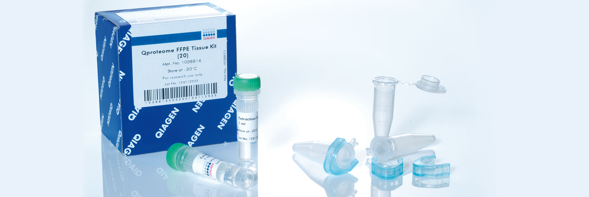
Yes. Supplementary protocols are available for the purification of full-length proteins from PAXgene Tissue fixed (PF) tissue and paraffin blocks using the Qproteome FFPE Tissue Kit (QIAGEN, cat. no. 37623). For more information, see the corresponding supplementary protocols under Resources.

9. Is it possible to microdissect PAXgene Tissue-fixed, paraffin-embedded (PFPE) and PAXgene Tissue-fixed, cryo-embedded (PFCE) tissues?
Yes, supplementary protocols for generating sections from PFPE and PFCE tissue blocks for manual and laser microdissection are available under Resources.

10. Which kits and protocols can be used for the isolation of nucleic acids from microdissected PAXgene Tissue-fixed, paraffin-embedded (PFPE) and PAXgene Tissue-fixed, cryo-embedded (PFCE) specimens?
Regular PAXgene Tissue kits can be used for the isolation of total RNA, including miRNA, and DNA from microdissected PFPE and PFCE tissue. Supplementary protocols developed specifically for the extraction of biomolecules from microdissected PFPE and PFCE samples are available under Resources.

Molecular analysis of biomolecules purified from PAXgene Tissue-treated samples

1. What is the RT-PCR performance of RNA purified from PAXgene Tissue-fixed, paraffin-embedded (PFPE) and PAXgene Tissue-fixed, cryo-embedded (PFCE) tissues compared to RNA from snap-frozen or formalin-fixed, paraffin-embedded (FFPE) tissues?
RNA, including miRNA, purified from PFPE and PFCE is free of chemical modifications and performs similarly or identically to RNA isolated from frozen tissue. For examples of the correlation of gene expression levels in snap frozen tissue, FFPE, and PFPE, see Figure 4 in Groelz et al. 2013 and Figure 3 in Viertler et al. 2012.

2. What is the PCR performance of DNA purified from PAXgene Tissue-fixed, paraffin-embedded (PFPE) and PAXgene Tissue-fixed, cryo-embedded (PFCE) tissues compared to DNA from snap-frozen or formalin-fixed paraffin-embedded (FFPE) tissues?
In contrast to FFPE, DNA purified from PFPE and PFCE is of high molecular weight and free of chemical modifications. In demanding downstream applications, such as multiplex or long-range PCR, it performs similarly or identically to DNA isolated from frozen tissue. For examples, see, Figure 5 in Viertler et al. 2012.

3. Is DNA purified from PAXgene Tissue-fixed, paraffin-embedded (PFPE) tissues suitable for targeted NGS analysis?
Yes. DNA purified from PFPE is of high molecular weight and free of chemical modifications. Several independent researchers successfully used DNA from PFPE for NGS with different sample types, workflows and platforms (data in preparation). The data confirm recent suggestions that some DNA sequence artifacts associated with FFPE can be avoided with PFPE (Högnäs et al. 2017).

4. How is the quality of proteins purified from tissues fixed and stabilized with the PAXgene Tissue System?
Proteins from PAXgene Tissue-fixed, paraffin-embedded (PFPE) and PAXgene Tissue-fixed, cryo-embedded (PFCE) tissues are non-degraded, immunoreactive and have been successfully investigated by western blot analysis, reverse-phase protein arrays, two-dimensional gel electrophoresis (2D-PAGE), enzyme-linked immunosorbent assay (ELISA) and MALDI imaging mass spectrometry.


Service

Contact us for troubleshooting and support

Global Contacts

Technical Service

Customer Care