Select your location:
Image of PAXgene Tissue DNA Kit (767134)

PAXgene Tissue DNA Kit

For isolation and purification of DNA from tissues fixed and stabilized using the PAXgene Tissue System

  • Effective purification of high-quality, unmodified DNA from tissue with preserved morphology
  • Protocols for paraffin-embedded or paraffin-free tissues
  • Automated purification on the QIAcube

Feature

Specification

Format

Spin column

Technology

Silica membrane

Main sample type

Human tissue

Sample amount

  • Up to 20 mg tissue fixed and (optionally) stabilized with PAXgene Tissue reagents
  • Up to 10 mg PF* tissue with high DNA content
  • 2–5 sections (5–10 µm thick) PFPE** tissue

Elution volume

14–40 µl

Time per run

30 min + 120 min incubation/8 samples

Processing

  • Manual: centrifugation
  • Automated: QIAcube

* PAXgene Tissue-fixed
** PAXgene Tissue-fixed, paraffin-embedded


Intended Use

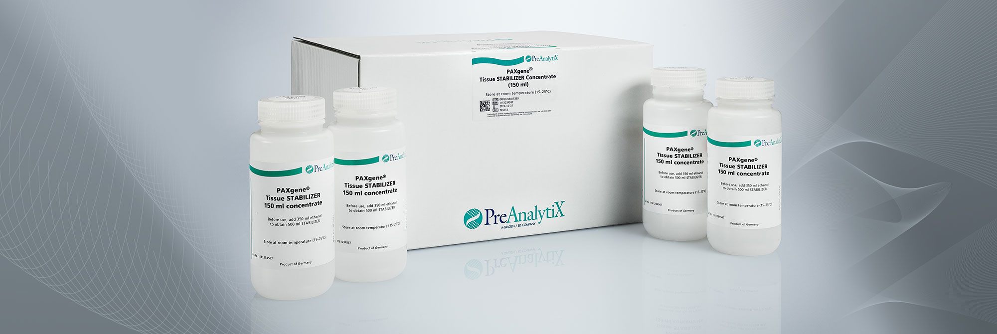
The PAXgene Tissue DNA Kit is intended for the purification of DNA from PAXgene Tissue-fixed (PF) and PAXgene Tissue-fixed, paraffin-embedded (PFPE) tissue samples. The kit is intended to be used as part of the PAXgene Tissue System, which additionally includes the PAXgene Tissue FIX Container and the PAXgene Tissue STABILIZER Concentrate for the collection, fixation, stabilization and storage of tissue samples.
 

For research use only. Not for use in diagnostic procedures. No claim or representation is intended to provide information for the diagnosis, prevention, or treatment of a disease. The performance characteristics of this product have not been fully established.

Order information

Product

Catalog No.

Price

PAXgene Tissue DNA Kit (50)

For 50 DNA preps: PAXgene DNA Mini Spin Columns, Processing Tubes, Microcentrifuge Tubes, Carrier RNA, and Buffers. To be used in conjunction with PAXgene Tissue FIX Containers + PAXgene Tissue STABILIZER reagent.

767134
359 USD
Order now
Order now

Details

Performance

Total DNA isolated with the PAXgene Tissue DNA Kit is highly pure. DNA has A260/A280 ratios of 1.7–1.9, and absorbance scans show a symmetrical peak at 260 nm, confirming high purity of genomic DNA. Contamination is minimal and purified DNA is ready to be used in downstream applications with no detectable inhibition of PCR (Figure 1 and Figure 2)

Principle

The PAXgene Tissue DNA Kit enables purification of DNA from tissues fixed and stabilized using the PAXgene Fixation and Stabilization reagents, which preserve tissue morphology and biomolecule integrity by avoiding destructive crosslinking and degradation found in formalin-fixed tissues. The purified DNA has no inhibitory chemical modifications and thus, can be used in sensitive downstream applications.

Procedure

PAxgene Tissue-fixed (PF) or PAXgene Tissue-fixed, paraffin-embedded (PFPE) tissue samples are lysed in Buffer TD1 with proteinase K. Conditions are adjusted for optimal binding of DNA to the silica membrane and the lysate is loaded on the PAXgene DNA spin column. DNA binds to the membrane while contaminants pass through. Enzyme inhibitors are effectively removed with subsequent washes and pure DNA is eluted in a low-salt elution buffer (Figure 3).

The PAXgene Tissue DNA Kit provides 2 protocols for purification of DNA from different starting materials:

  • Sections of PFPE tissue
  • PF tissue samples (without paraffin embedding)

Automation

DNA purification using the PAXgene Tissue DNA Kit can be automated on the QIAcube, enabling:

  • Elimination of manual processing steps
  • Elimination of up to 75% hands-on time compared to manual processing (Figure 4
  • Processing of up to 12 samples per run
  • Processing of normal, fibrous or fat-rich PAXgene Tissue-fixed samples (Figure 5)
  • Processing of 2–5 sections of PFPE tissue samples (Figure 8)

Two protocols are available for automated purification from different starting materials:

Applications

The purified DNA is ready to use in a wide range of downstream applications (see Resources), including:

  • PCR, multiplex, long-range, and quantitative, real-time PCR
  • Southern blotting
  • SNP genotyping
  • Next-generation sequencing (NGS)

Resources

PAXgene Tissue DNA Kit Handbook

703.1 KB

Download

Deparaffinization of PAXgene Tissue-fixed, Paraffin-embedded Tissue (PFPE) Sections with Deparaffinization Solution

614.1 KB

Download

Simultaneous Purification of Genomic DNA and Total RNA, including miRNA, from Sections of PAXgene Tissue-fixed, Paraffin-embedded (PFPE) Tissue

82.6 KB

Download

Purification of Genomic DNA from Microdissected PAXgene Tissue-fixed, Paraffin-embedded (PFPE) and PAXgene Tissue-fixed, Cryo-embedded (PFCE) Tissues

699.3 KB

Download

Purification of Genomic DNA from Sections of PAXgene Tissue-fixed, Cryo-embedded (PFCE) Tissue Placed Directly Into a Microcentrifuge Tube

589.0 KB

Download

PAXgene Tissue System Brochure

1.9 MB

Download

DNA Isolation with the PAXgene Tissue DNA Kit

1.9 MB

Download

(EN) Important Note: PreAnalytiX GmbH street address has changed from “Feldbachstrasse” to “Garstligweg 8”

158.9 KB

Download

Detection of PI3K mutational status in DNA from human breast cancer PFPE tissue using the PI3K Mutation Test Kit (QIAGEN®)

1.2 MB

Download

Quantitative Analysis of KRAS and BRAF Mutational Status in DNA from PAXgene Tissue-fixed, Paraffin-embedded (PFPE) Tissue Using Pyrosequencing Technology

1.3 MB

Download

MSDS PAXgene Tissue DNA Kit

Learn more

DNA Quality Measurement and Somatic Mutation Profiling in PAXgene Tissue Samples with qBiomarker Somatic Mutation PCR Arrays (Long 2012)

1.2 MB

Download

PAXgene Tissue Fixation Technology for Simultaneous Preservation of Morphology and Biomolecules (Groelz 2012)

1.2 MB

Download

Preservation of Morphology and Biomolecules Within Tissue Stored for Three Years at –80°C in PAXgene Tissue Stabilizer Reagent (Groelz 2012)

890.5 KB

Download

Morphological, Epigenomic and Mutational Analyses of PAXgene Tissue-fixed, Paraffin-embedded (PFPE) Colorectal Cancer (CRC) Specimens — Comparison to Formalin-fixed, Paraffin-embedded (FFPE) and Snap-frozen Samples (Groelz 2010)

1.1 MB

Download

New Fixation Technology for Simultaneous Preservation of Mophology and Nucleic Acids in Tissue (Groelz 2008)

516.6 KB

Download

PAXgene Tissue: A New TIssue Fixation Technology for Simultaneous Preservation of Morphology and Nucleic Acids (Groelz 2011)

542.1 KB

Download

Nurminen et al.(2023) Cancer origin tracing and timing in two high-risk prostate cancers using multisample whole genome analysis: prospects for personalized medicine. Genome Medicine volume 15, Article number: 82

Learn more

Ellis, P. et al. (2021) Reliable detection of somatic mutations in solid tissues by laser-capture microdissection and low-input DNA sequencing. PubMed.gov.

Learn more

Wiethaler, M. et al. (2019) BarrettNET – a prospective registry for risk estimation of patients with Barrett's esophagus to progress to adenocarcinoma. Dis. Esophagus 32: doz024. doi: 10.1093/dote

Learn more

Liu, Y. and Edward, D.P. (2017) Assessment of PAXgene Fixation on Preservation of Morphology and Nucleic Acids in Microdissected Retina Tissue. Curr. Eye Res. 42, 104. E-published in 2016. doi: 0.3

Learn more

Ruusuvuori, P. et al. (2016) Feature-based analysis of mouse prostatic intraepithelial neoplasia in histological tissue sections. J. Pathol. Inform. 7, 5. doi: 10.4103/2153-3539.175378

Learn more

Loibner, M. et al. (2016) Pathogen Inactivating Properties and Increased Sensitivity in Molecular Diagnostics by PAXgene, a Novel Non-Crosslinking Tissue Fixative. PLoS One 11, e0151383. doi: 10.1

Learn more

Oberauner-Wappis, L. et al. (2016) Protocol for HER2 FISH determination on PAXgene-fixed and paraffin-embedded tissue in breast cancer. Int. J. Exp. Pathol. 97, 202. doi: 10.1111/iep.12185

Learn more

Korenkova, V. et al. (2016) The focus on sample quality: Influence of colon tissue collection on reliability of qPCR data. Sci. Rep. 6, 29023. doi: 10.1038/srep29023

Learn more

Mathieson, W. et al. (2016) A Critical Evaluation of the PAXgene Tissue Fixation System:  Morphology, Immunohistochemistry, Molecular Biology, and Proteomics. Am. J. Clin. Pathol. 146, 25. doi: 0.

Learn more

Gillard, M. et al. (2015) Next-gen tissue: preservation of molecular and morphological fidelity in prostate tissue. Am. J. Transl. Res. 7, 1227.

Learn more

Groelz, D. et al. (2018) Impact of storage conditions on the quality of nucleic acids in paraffin embedded tissues. PLoS ONE 13(9): e0203608.

Learn more

PAXgene Tissue DNA Kit

Purification and quality of biomolecules from PAXgene Tissue-treated samples

1. Is it possible to use formalin-fixed, paraffin-embedded (FFPE) kits and protocols to isolate biomolecules from PAXgene Tissue-fixed, paraffin-embedded (PFPE) tissues?
No. Procedures developed for the extraction of biomolecules from FFPE tissues include prolonged proteinase K digestion and heating steps to remove chemical modifications introduced by formalin. Since the PAXgene Tissue System does not chemically modify biomolecules, these steps are not necessary and, in fact, lead to degradation of biomolecules. Instead, use dedicated PAXgene Tissue Kits and supplementary protocols for extraction of biomolecules from PAXgene Tissue-treated samples.


2. What is the purity of nucleic acids extracted with the PAXgene Tissue Kits?
The PAXgene Tissue DNA and RNA, including miRNA, Kits are based on proven QIAGEN technologies. Nucleic acids isolated with these kits are generally of high purity.

On average, measurements of the A260/A280 ratio for DNA purified with the PAXgene Tissue DNA Kit are >1.7, and ratios for RNA, including miRNA, purified with the PAXgene Tissue RNA/miRNA Kit are >1.8.

3. How well is DNA integrity preserved in PAXgene Tissue-fixed, paraffin-embedded (PFPE) tissues?  
In contrast to DNA isolated from formalin-fixed, paraffin-embedded (FFPE) tissue, DNA from PFPE tissue exhibits high molecular weight. In most cases, a distinct 10 kb band is observed in electrophoretically separated DNA eluates. For an example, see Figure 2 in the Technical Note "Quantitative analysis of KRAS and BRAF mutational status in DNA from PAXgene Tissue-fixed, paraffin-embedded (PFPE) tissue using Pyrosequencing technology" under Resources.   

4. What are the yield and integrity of nucleic acids isolated from blocks of PAXgene Tissue-fixed, cryo-embedded (PFCE) tissues?    
DNA and RNA isolated from PFCE tissue specimens are of high quantity and quality, comparable to PFPE tissue.    


5. Are special kits and protocols required for the isolation of biomolecules from PAXgene Tissue fixed, cryo-embedded (PFCE) tissues?    
No. Regular PAXgene Tissue Kits can be used for the isolation of RNA, miRNA and DNA from PFCE tissue. Supplementary protocols developed specifically for the extraction of biomolecules from PFCE samples are available under Resources.


6. Which kits and protocols can be used for the isolation of nucleic acids from microdissected PAXgene-Tissue fixed, paraffin-embedded (PFPE) and PAXgene Tissue-fixed, cryo-embedded (PFCE) specimens?    
Regular PAXgene Tissue Kits can be used for the isolation of RNA, miRNA and DNA from microdissected PFPE and PFCE tissue. Supplementary protocols developed specifically for the extraction of biomolecules from microdissected PFPE and PFCE samples are available under Resources.    


Molecular analysis of biomolecules purified from PAXgene Tissue-treated samples    

1. What is the PCR performance of DNA purified from PAXgene Tissue-fixed, paraffin-embedded (PFPE) and PAXgene Tissue-fixed, cryo-embedded (PFCE) tissues compared to RNA from snap-frozen or formalin-fixed, paraffin-embedded (FFPE) tissues?    
In contrast to FFPE, DNA purified from PFPE and PFCE is of high molecular weight and free of chemical modifications. In demanding downstream applications, such as multiplex or long-range PCR, it performs similarly or identically to DNA isolated from frozen tissue. For examples, see, Figure 5 in Viertler et al. 2012  under Resources.

2. Is DNA purified from PAXgene Tissue-fixed, paraffin-embedded (PFPE) tissues suitable for targeted NGS analysis?
Yes. DNA purified from PFPE is of high molecular weight and free of chemical modifications. Several independent researchers successfully used DNA from PFPE for NGS with different sample types, workflows and platforms (data in preparation). The data confirm recent suggestions that some DNA sequence artifacts associated with FFPE can be avoided with PFPE (see Högnäs et al. 2017 under Resources).

Service

Contact us for troubleshooting and support

Global Contacts

Technical Service

Customer Care